Sep 27, 2022 | General Chiropractic
Tumor and spine — two words we do not relish hearing in the same sentence. When speaking about medical conditions, both the spine and tumors are not to be taken lightly. Whether benign or malignant, a tumor can cause pain and menace your daily life. There are several different types of and causes for spinal tumors, but each one requires professional medical attention.
What is a Spinal Tumor?
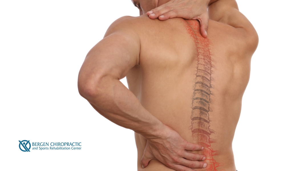
To understand spinal tumors, one must first understand the basic structure of the spine. The long stack of bones along your back is called the backbone or spine. A flexible column, the spinal cord encases the spinal column, protecting it from daily impact. Running from the base of the skull to the end of your tailbone, the backbone safeguards nerve bundles and cells in the central nervous system responsible for delivering messages from the brain and spinal cord to the rest of the body (or vice versa). A spinal tumor is so named because it is located within or near the spinal cord or spinal column. Spinal tumors are abnormal tissue masses with cells that uncontrollably grow and multiply. There are two primary spinal tumor types:
- Benign tumors or non-cancerous;
- Malignant tumors or cancerous.
Spinal tumors are further categorized as:
- Primary spinal tumors: They stem from the spine or spinal cord.
- Metastatic tumors (secondary tumors): They develop after cancer has spread from a different site to the spine.
While the exact cause of most primary spinal tumors is still largely considered a mystery, there are some known causes and connections such as:
- Exposure to cancer-causing agents;
- Compromised immune systems;
- Genetics;
- Blood vessel connection.
Compromised immune systems can result in certain primary spinal tumors such as spinal cord lymphomas, which attack immune cells called lymphocytes. Genetics and neurological disorders can also contribute to one’s propensity to develop primary spinal tumors. For example, neurofibromatosis 2 is hereditary and begins as benign lesions that develop on or near nerves related to hearing. Extensive neurofibromatosis 2 can lead to hearing loss in one or both ears, and those suffering from this disorder are also known to develop spinal canal tumors. Finally, some primary spinal tumors are connected to blood vessels. For example, von Hippel-Lindau disease is a rare multisystem disorder that causes tumors in the blood vessels connected to the brain, retina, or spinal cord. Other primary spinal tumors in the kidneys or adrenal glands also result from this spinal disease.
What are the Different Spinal Cord Tumors?
There are four different types of spinal cord tumors, categorized by the spinal region they affect:
- Cervical, which affects the neck area;
- Thoracic, which affects the upper to mid back;
- Lumbar, which affects the lower back;
- Sacrum, which affects the bottom of the spine.
Spinal cord tumors are further classified as one of the following based on their location within the spine:
- Intradural-extramedullary;
- Intramedullary;
- Extradural.
Intradural-Extramedullary Tumors
An intradural-extramedullary tumor is located within the thin covering or dura of the spinal cord but is still outside the actual spinal cord. Tumors are found within this area about 40% of the time and are either in the spinal cord’s arachnoid membrane, in the spinal cord nerve roots which branch out from the spinal cord, or at the base of the spinal cord. While tumors in the arachnoid membrane are frequently benign, they are also challenging to remove and may reappear. Also, often benign, nerve root tumors may become malignant through time. Finally, the tumors at the spinal cord base can be large, causing difficulty removing them from the more delicate and fine neural structures.
Intramedullary Tumors
Intramedullary tumors grow inside the spinal cord and typically stem from glial cells found throughout the spinal cord’s interstitium. About 5% of tumors occur in this region. Astrocytomas and ependymomas are two of the most common types of intramedullary tumors. Also generally benign, these tumors can be difficult to remove.
Extradural Tumors
Finally, extradural tumors are found outside of the dura. Tumors grow in this area approximately 55% of the time. Typically, these tumors come from metastatic spinal cancer or, less commonly, from schwannomas from cells that cover nerve roots. Sometimes, extradural tumors can poke through the intervertebral foramina, causing it to lie between the inside and outside of the spinal canal.
What Percentage of Spinal Tumors are Benign?
While we all wished that osseous spinal tumors were always benign, that is not always the case. There are plenty of different types of spinal tumors that are benign, such as:
- Giant cell bone tumors;
- Osteochondroma;
- Hemangioma.
Spinal osseous tumors occur in any part of the vertebrae, most often in the thoracic vertebrae or the sacral and lumbar spine — they rarely occur in the cervical spine. Those who are middle-aged or elderly are more likely to develop malignant spinal tumors. On the other hand, younger patients are more prone to benign tumors. Most spinal tumors are secondary or metastatic spinal tumors. Almost 97% of all spinal tumors are metastatic, and about 10,000 people in the United States are diagnosed yearly with metastatic spinal cord tumors. Approximately 30% to 70% of cancer patients will develop cancer metastasis in their spine. More uncommon, benign primary spinal tumors make up about 0.5% of all newly diagnosed tumors. However, malignant primary spinal tumors are considered even more uncommon.
Spinal Tumor Early Symptoms
The type of symptoms for early diagnosis develops based on the following factors:
- The tumor location within or along the spinal column;
- The tumor size;
- The type of tumor.
However, symptoms sometimes fail to develop based on the tumor size! If the tumor is small and does not pressure or irritate nearby tissues, a patient will probably not experience any symptoms. However, once it increases in size, the tumor can cause different painful or uncomfortable symptoms along the spinal column. The most common symptom of either a benign or malignant spinal tumor is back pain and muscle weakness. Tumors are more likely to develop in the spine’s middle or lower back regions. As such, the pain will often target these spinal areas. While back pain is not uncommon in more elderly or particularly active individuals, pain from a spinal tumor has some unique attributes. Pain caused by a spinal tumor is not specific to a previous injury or physical activity but can increase from exercising, sneezing, or coughing. Spinal tumor pain is also deep and aching, starting slowly and increasing gradually. At night, the pain can be increasingly persistent and disrupt sleep. As the tumor grows, the pain can become intense and cause disruption even while resting. Radicular pain spreads from the spinal column to the hips, legs, feet, or arms and is a painfully sharp and shooting symptom of spinal tumors. If the spinal tumor is in a specific location and grows to press on the spinal cord or nerve roots, blood vessels, or bones of the spine, then other symptoms such as the following may develop:
- Numbness, tingling, or loss of sensation in your legs, arms, or chest;
- Leg, arm, or chest muscle weakness;
- Muscle cramps or spasms;
- A stiff back or neck;
- Loss of bowel or bladder function;
- Difficulty walking;
- Scoliosis or a different spinal deformity resulting from a large, destructive tumor growth;
- Varying degrees and locations of paralysis, depending on the spinal nerves the tumor is compressing.
Diagnosing Tumors
There are three main methods for diagnosing tumors:
- Spinal magnetic resonance imaging tests (MRI);
- Computerized tomography (CT scan);
- Biopsy.
Magnetic Resonance Imaging (MRI)
A magnetic resonance imaging or MRI is a special device that uses powerful magnetic fields and radio waves to create accurate images of your spine, spinal cord, and nerves. The preferred testing method, an MRI enables professionals to view the spinal cord and surrounding soft tissue. While some may become claustrophobic within an MRI scanner or find the loud thumping sound disturbing, your health care professionals have methods of making the experience more comfortable. Whether you use earplugs or a distracting device such as a TV or headphones, there are many different ways to overcome anxiety regarding your MRI. Sometimes, you may even be given a general anesthetic to make the process more comfortable.
Computerized Tomography (CT Scan)
Computerized tomography (CT scan) employs a narrow radiation beam to form detailed spinal images. It is sometimes combined with an injected contrast dye to make any abnormal growth in the spinal canal or spinal cord even clearer. However, unlike an MRI, a CT scan is rarely used to help diagnose a spinal tumor.
Biopsy
Finally, a biopsy is the only method to determine the specific type of spinal tumor you are dealing with. By taking a small soft tissue sample or biopsy and examining it under a microscope, a biopsy result can help determine the best spinal cancer treatment options for your situation.
Can Spinal Tumors Be Removed?
The good news is that spinal tumors can be removed! Radiation therapy, chemotherapy, or spinal surgery are three methods by which to evict these pesky tenants.
Radiation Therapy
Although radiation therapy sounds scary, the basic process is actually rather simple. By using high doses of x-rays, this treatment for spinal tumor cells destroys or shrinks the tumor body. Once the tumor is small enough, the pain and other symptoms are reduced or eliminated.
Chemotherapy
Chemotherapy is a standard method to destroy spinal cancer cells. By taking chemotherapy drugs, any cancer cells found in your spine tumors will be eliminated. Either through injection into a vein or by ingesting a pill, healthcare providers use chemotherapy to shrink tumors before surgery or to kill them completely.
Spinal Surgery
Spinal surgery is generally reserved for those with metastatic spinal tumors when they are estimated to live three to four months or longer, and the tumor is not responding to radiation or chemotherapy. Pain and other symptoms can be relieved when the spine is stabilized, and the neurological function is preserved. Neurological surgeons perform either traditional open surgery or a minimally-invasive procedure. It depends on the opinion of your spinal surgeons to perform vertebroplasty or kyphoplasty. Both of these procedures provide spinal cord support and stability, relieving pain and improving mobility. About 10% of patients with symptomatic spinal metastases are eligible for surgical treatment.
How Can Chiropractic Care Help with Spinal Tumors?
Chiropractic care can be useful in controlling pain, headaches, and tension. Some scientific evidence suggests that chiropractic treatment options may also relieve headaches and back pain. However, there is no substantial evidence that chiropractic treatment can prevent, treat, or cure spinal cancer. Thus, while it is a useful tool to manage painful symptoms, it is not at all a miracle cure for spinal tumors.
Do You Have More Questions Regarding Spinal Tumors? Dr. Doerr Can Help!
At the Bergen Chiropractic and Sports Rehabilitation Center, our chiropractic team, led by Dr. Gregory Doerr, follows the highest and most professional medical standards to provide superior chiropractic help. After all, our mission is to provide unparalleled patient care and services in a comfortable healing atmosphere. Access our contact form or call us at (201) 298-7179 or (201) 357-6539 to learn more about our chiropractic services! Our chiropractic offices at 532 Anderson Avenue, Cliffside Park, NJ 07010, and 62 Summit Ave, Hackensack, NJ 07601 are ready to welcome you as we proudly serve New York, New Jersey, Philadelphia, PA, and Baltimore, MD areas. Also, feel free to access our blog page and social media for more information on chiropractic treatments!
Sep 20, 2022 | General Chiropractic
Scoliosis is perhaps one of the better-known health conditions which affect the spine. Sometimes causing pain and nerve damage, more severe cases of scoliosis can make everyday tasks difficult. Thankfully, there are solutions to help those suffering from adult scoliosis. Whether through physical therapy management or surgery, science and medicine have found ways to aid people with scoliosis. Continue reading to learn more about adult scoliosis, its symptoms, and treatment options!
What is Adult Scoliosis?
While it is natural and healthy for the spine to have a forward-and-backward curve, scoliosis is a condition that causes the spine to curve beyond its healthy limits. An abnormal curvature of the spine, scoliosis causes the spine to rotate and develop a side-to-side curve. Scoliosis can cause a curve as mild as 10 degrees or as severe as over 100 degrees!
What are the Main Scoliosis Types?
There are four main types of scoliosis that progress in relatively similar fashions:
- Congenital scoliosis;
- Neuromuscular scoliosis;
- Adolescent idiopathic scoliosis;
- Adult de novo scoliosis.
Congenital, neuromuscular, and idiopathic scoliosis are primarily diagnosed in children. The most common type of adult scoliosis, degenerative scoliosis, curves the spine with age. Luckily, spinal curvatures are often mild cases and do not require treatment.
What is Causing My Adult Scoliosis?
Adult scoliosis stems from various causes depending on its type. Scoliosis without a known cause, or idiopathic scoliosis, is often uncovered during childhood or adolescence growth. Adult scoliosis can also be a case of pediatric scoliosis, left undiscovered until adulthood. However, it is more common for adult scoliosis symptoms to develop through aging and degenerative changes.
There are four types of scoliosis that adults can develop:
- Degenerative scoliosis is found in older individuals whose spinal discs have deteriorated with the natural aging process. Aging can thus lead to the compression of the vertebral column and the development of abnormal spinal curvature.
- A little more mysterious, traumatic scoliosis is somehow caused by severe trauma or accidents. How a spinal deformity develops in such a manner is still unknown.
- Pathological scoliosis is a result of tumors pressing on the spine. Spinal tumors can exert such a strong pressure on the spine that a curvature develops.
- As mentioned earlier, idiopathic scoliosis represents cases of undiagnosed adolescent scoliosis. Instances of such conditions are rare and difficult to confirm or track.
Common Adult Scoliosis Symptoms
Scoliosis-related symptoms are not actually caused by the curvature of the spine, although they are connected with the degeneration of the spine as time passes. Such symptoms are treated in a similar fashion, despite the actual diagnosis of scoliosis.
Although most cases of adult scoliosis are symptomless, back or leg pain is a common sign of this disease. Back pain can result from multiple medical conditions, including:
- Arthritis;
- The inability to stand upright;
- Weakness of the core musculature.
Pain, numbness, or weakness in the legs are common symptoms of spinal stenosis, which occurs if there is tremendous pressure on the nerves in the lumbar spine.
Sometimes, body changes such as the following may develop:
- Height loss;
- Shortness of breath or quick fatigue;
- Uneven alignment of the pelvis and hips.
Depending on the degree of the spinal curvature, internal symptoms may be accompanied by changes in the body’s outward appearance. For example, a bulge or spinal deformity can become visible due to muscle or rib cage rotation. An inability to stand upright might also be a symptom.
How is Adult Scoliosis Diagnosed?
The first step toward diagnosis and treatment is for your healthcare professional to examine you and your family’s medical history. Some common diagnostic questions circle around:
- Family history;
- When you first noticed the spinal changes;
- Curve progression;
- Presence and location of any pain;
- Bowel, bladder, or motor dysfunction.
Next, wellness professionals will perform a physical examination and medical evaluation to check the shape of your spine curves and your movement. They may also use reflex, sensation, and muscle-strength checks to test your nerves.
After a physical exam, your doctor will recommend X-rays to comprehensively inspect your spine’s front and side. With the help of such imaging, your doctor can determine the type and severity of your scoliosis.
What are My Non-Operative Treatment Options?
For adults diagnosed with scoliosis, the extent of the spinal curve may not necessarily determine the treatment options. Instead, treating scoliosis stems from the goal of relieving symptoms and not necessarily curing the curve.
As such, many patients with adult scoliosis conditions that are not severe cases are treated through noninvasive means, such as:
- Regular medical observation;
- Over-the-counter pain medications;
- Core-strengthening exercises to improve abdomen and back flexibility.
Scoliosis Exercises
Often, your doctor will recommend using physical therapy to maintain strength and relieve pain. A physical therapist may help improve scoliosis by:
- Working to improve posture;
- Doing low-impact exercises;
- Daily stretching;
- Staying active.
Arm/Leg Raise
Arm/leg raises are useful to strengthen your lower back and core muscles, supporting your spine. To perform this exercise:
- Start by lying on your stomach with your chin or forehead on the ground.
- With your legs straight and arms extended overhead, slowly raise one arm off the ground, hold it for a moment, and then slowly lower it back to the ground.
- Repeat these steps for your opposite leg and arm with 15 repetitions each.
Pelvic Tilt
Pelvic tilts strengthen your abdominal muscles. Be sure to perform them on a yoga mat for support:
- First, lie on your back with your knees bent and both feet flat on the floor, toes pointed forward.
- Pull in your belly button so your pelvis pushes toward the ceiling and your back flattens against the ground.
- Hold the position for 20 seconds, and then slowly relax.
- Repeat the exercise 10 times.
Cat/Cow Stretch
Cat/cow stretch focuses on the muscles and tendons which support your spine:
- Start by getting on your hands and knees. Keep your arms straight under your shoulders and your knees under your hips.
- While looking at the floor, keep your head straight in line with your torso and spine.
- Now, round your back and lift your spine toward the ceiling. At this point, your eyes should face your belly.
- Hold the position and take a deep breath before slowly lifting your chest and tailbone toward the ceiling. Let your stomach sink toward the ground as your eyes look up toward the ceiling.
- After another deep breath, slowly round your back and lift your spine toward the ceiling.
- Continue alternating between the poses to gently stretch and strengthen your abdominals and lower back.
Latissimus Stretch
Finally, the latissimus stretch loosens and strengthens tight lat muscles:
- To begin, stand with your feet shoulder-width apart and your knees slightly bent.
- Now, reach overhead and grab your left wrist with your right hand.
- Next, with most of your body weight on your right leg, bend at your right side until you feel a gentle stretch along your left trunk.
- Hold the position for 5 to 10 seconds before returning to the start position by pushing from your right foot.
- Be sure to repeat the exercise on your left foot and the opposite side for maximum benefits.
Invasive Treatment Options for Adult Scoliosis Patients
If oral medications or physical therapy are inadequate for reducing pain, your doctor may recommend epidurals around the spinal cord or nerve block injections for symptom relief.
Surgery, another option for severe scoliosis, is considered a last-resort avenue because of the risks associated with spinal surgery.
Spinal Injections
Sometimes, scoliosis can irritate or pressurize the nerves in and around the spine, causing pain, numbness, and tingling in your lower back down to your feet. If this is the case, spinal injections such as steroids and local anesthetic may offer some relief. Unfortunately, the benefits of such injections last for only a few weeks or months and are not a long-term solution.
Back Braces
Not widely used in adults with scoliosis, back braces are occasionally helpful in providing pain relief through spinal support. A brace may be an alternative to surgery if you are not well enough to undergo such an operation.
Surgical Treatment Plan
While it is not required for most patients with adult scoliosis, lumbar decompression surgery may be considered for the following reasons:
- If the curve in your spine is severe or significantly worsening;
- If you have spinal imbalance;
- If you have severe back pain and other treatments have not helped;
- If your spinal nerves are being irritated or squashed.
Should surgery be deemed necessary, there are three different surgical techniques:
- Laminectomy;
- Discectomy;
- Spinal fusion.
A laminectomy procedure removes one section of bone in your spinal canal to relieve nerve pressure. The second option, a discectomy, is where one section of discs between the vertebrae is removed to relieve pressure. Thirdly, a spinal fusion takes two or more vertebrae and joins them together to stabilize, strengthen, and straighten the spine. Oftentimes, a combination of these techniques is used instead of just one.
A posterior spinal fusion is the most common type of special surgery for adults with scoliosis. For this procedure, the orthopedic surgeon makes an incision from the back, places screws to collect scoliosis, and then ‘welds’ the vertebrae together with bone chips. The surgeon may take these chips from elsewhere in the patient’s own body or from a donor through a bone bank.
It may take six months to over one year for the healing process to be complete. However, recovery from spinal surgery only takes four to six weeks. During the recovery period, rods, screws, or other implants may be used to hold the spine in alignment. After the fusion is complete, the implants are no longer necessary but are left within the patient to avoid additional surgery. Normal mobility is achievable after healing if the treatment primarily targets the thoracic spine. However, treatment in the lumbar spine will result in more restricted sideways movement.
Dr. Doerr is the Best Chiropractor in New Jersey for Treating Adult Scoliosis!
At the Bergen Chiropractic and Sports Rehabilitation Center, our chiropractic team, led by Dr. Gregory Doerr, follows the highest and most professional medical standards to provide superior chiropractic help. After all, our mission is to provide unparalleled patient care and services in a comfortable healing atmosphere. Access our contact form or call us at (201) 298-7179 or (201) 357-6539 to learn more about our chiropractic services! Our chiropractic offices at 532 Anderson Avenue, Cliffside Park, NJ 07010, and 62 Summit Ave, Hackensack, NJ 07601 are ready to welcome you as we proudly serve New York, New Jersey, Philadelphia, PA, and Baltimore, MD areas. Also, feel free to access our blog page and social media for more information on chiropractic treatments!
Sep 13, 2022 | General Chiropractic
It can be challenging to understand spinal fractures, as they all have different causes, symptoms, and treatments. Whether in the thoracic or lumbar spine, fractured vertebrae can lead to further injury if not dealt with immediately. There are several pain management and treatment options for spinal fractures, be it through physical therapy or surgical procedure.
What is a Vertebral or Spinal Fracture?
A spinal injury can range from a mild ligament or muscle strain to fractures or dislocations of the bony vertebrae. When a vertebra becomes seriously fractured or dislocated, it can cause bone fragments to pinch and damage the spinal nerves or spinal cord! Many spinal injuries do not require surgery; however, severe long-term issues can result from untreated or mistreated major fractures.
Most spinal fractures occur in the lower back region — 64% occur in the thoracolumbar or lower back region, while 5-10% occur in the cervical or neck region.
How Do Spinal Fractures Occur?
The spine is rather delicate as even minor falls or trauma can produce spine fractures. The primary causes of spinal fractures are:
- Car accidents (45%);
- Falls (20%);
- Sports (15%);
- Acts of violence (15%);
- Various activities (5%).
Other causes of spinal fractures are diseases such as spine tumors and osteoporosis, which weakens the bones, leading to severe pain and vertebral compression fractures.
Spinal Fracture Symptoms
There are various symptoms of a spinal fracture that depend on the severity and location of the injury. Some symptoms include:
- Back or neck pain;
- Numbness;
- Tingling;
- Muscle spasm;
- Weakness;
- Bowel/ bladder changes;
- Paralysis or loss of movement in the arms or legs.
Paralysis can indicate a more profound spinal cord injury. Luckily, fractures do not always cause spinal cord damage, and rarely is the spinal cord completely severed.
How are Spinal Fractures Diagnosed?
Often, a spinal injury will land you in the emergency room, and an Emergency Medicine specialist will be the first to examine the injury. For severe fractures, other specialists will be called in for further assessment. Doctors will check your breathing and perform a physical exam of the spine. Until adequately diagnosed, the spine is kept immobile in a neck or back brace.
CT Scan
The only way to properly diagnose the extent of a spinal injury is through an X-ray, which views the bony vertebrae in your spine, revealing any fractures. A Computed Tomography (CT) scan is the next step toward diagnosis. Safe and noninvasive, a CT scan uses an X-ray beam and a computer to create two-dimensional images of the spine, helpful in viewing any abnormal movement in the bony structures.
MRI
Finally, a Magnetic Resonance Imaging (MRI) scan is another noninvasive test that uses magnetic fields and radiofrequency waves to detail the soft tissues of the spine. An MRI offers visibility of the nerves and discs, allowing evaluation of soft tissue damage to ligaments and discs.
How are Vertebral Fractures Classified?
Vertebral fractures are classified as either:
- Stable and Unstable Fractures;
- Minor and Major Fractures;
- Denis Classification Method.
Each classification type differentiates the injury’s severity, placement, and stability. Together, they can help doctors determine the best treatment option for vertebral fractures.
Stable and Unstable Fractures
A stable fracture does not cause spinal deformity or neurological (i.e., nerve-related) problems, permitting your spine to carry and distribute weight — of course, not as well as if there were not a fracture, but well enough to function securely.
On the other hand, unstable fractures make it difficult for the spine to carry or distribute weight. Progressing and causing further damage, these fractures can result in spinal deformity.
Minor and Major Fractures
A minor fracture occurs when part of the vertebra’s posterior or back side elements has broken. Minor fractures affect those parts of the spine that are less important to spinal column stability.
For example, the spinous process and facet joints are the posterior elements of the spine. The injury is not considered overly serious when fracturing this part of the vertebrae.
Major fractures are defined as the fracture of the pedicles, lamina, or vertebrae. Vertebral body fractures are considered major because these areas carry so much weight and are responsible for distributing force. When these parts of the spine fracture, they cause severe vertebral misalignment and increase the risk of nerve damage. Because the pedicles and lamina are necessary to keep your spine stable, your spine becomes unstable when they fracture.
The Denis Classification
The Denis Classification method is commonly utilized by doctors to classify a spinal fracture in the thoracic, lumbar, or thoracolumbar spinal regions. Doctors can efficiently classify a fracture’s severity and placement by dividing the lateral or side spine into three sections (i.e., front, middle, and back).
The anterior column is the front part of the vertebra or the part that faces in towards your body. Half of the front anterior column is the vertebral body and intervertebral disc.
Next, the middle column or the back half of the vertebral body is vital for spinal stability. When a fracture occurs in the middle and anterior or posterior columns, nerve damage and spinal instability are likely. On the other hand, if the middle column remains strong and intact, you have an increased chance of stability.
Finally, the back column includes all sections of the vertebra on the back side of the posterior column. The pedicles, lamina, and spinous process are all part of the posterior column.
Visualizing three columns makes diagnosing the extent of a spinal fracture and its stability easier. For example, if the fracture only affects the front or anterior column, the spine may be sufficiently stable to withstand the body’s weight. However, unlike posterior fractures, spinal fractures in the anterior or middle column are generally unstable.
Types of Vertebral Fractures
There are four main types of vertebral fractures:
- Compression;
- Burst;
- Flexion-distraction;
- Fracture dislocation.
#1: Compression Fracture
Compression fractures are common in patients with osteoporosis or other bone-weakening diseases. While the vertebra can absorb a lot of pressure, there is only so much it can handle before the bone fractures under stress. A sudden force or tremendous pressure can cause vertebrae to fracture.
Is a Compression Fracture the Same As a Broken Back?
When a bone sustains more pressure than it can handle, it fractures or breaks. A vertebral compression fracture is the most common vertebral fracture and occurs when a downward force shatters or collapses the vertebrae. A large enough force may even send bone fragments into the spinal canal, resulting in a burst fracture. In this way, a more severe spinal cord compression fracture is essentially the same as a broken back.
#2: Burst Fracture
Caused by severe trauma such as a car accident, burst fractures happen when an extreme force crushes the vertebra. A burst fracture not only affects one part of the vertebra but fractures it in several places. In addition, bony fragments spread throughout when the vertebra is crushed so wholly, causing spinal cord injury.
#3: Flexion-distraction Fracture
A flexion-distraction fracture results after severe trauma. For example, if your spine is forced to flex forward while another sudden forward movement places incredible stress on the spine, your vertebrae will break.
#4: Fracture Dislocation
The final type of vertebral fracture is fracture dislocation, a double doozy spinal injury. You are suffering from fracture dislocation when you experience any of the above fractures and the vertebra moves significantly or dislocates. Generally, these fractures involve all three columns of the Denis Classification, causing extreme spinal instability.
How are These Fractures Treated?
Treatment for vertebral fractures depends on:
- Other injuries and their treatment;
- The particular fracture pattern;
- Whether a neurological injury is present or not.
After stabilization of other life-threatening injuries, the doctor evaluates the spinal fracture pattern to decide if spine surgery is necessary.
A stable fracture may not require surgical treatment but can be stabilized by wearing a neck or back brace for 8 to 12 weeks. On the other hand, an unstable neck fracture or dislocation may require more treatment through traction, which realigns the spine to its correct position.
Spine surgery is often required for unstable burst fractures with:
- Multiple bone fragments;
- Severe loss of vertebral body height;
- Excessive forward bending near the injury;
- Significant nerve injury or disk pinching;
- Unstable ligament damage.
Flexion fracture patterns with the above symptoms are treated with surgical decompression of the spinal canal and stabilization of the fracture. A laminectomy surgically decompresses the spine by removing the bony arch or structure, pressuring the spinal cord on the backside of the spinal canal. It relieves pressure by providing extra space for the spinal cord to drift backward.
Fusion, another surgical treatment used to treat unstable fractures, joins two vertebrae with a bone graft and hardware such as plates, rods, or cages and transforms two vertebrae into a solid piece of bone.
Minimally invasive procedures also treat vertebral compression fractures commonly caused by osteoporosis and spinal tumors. For example, vertebroplasty injects bone cement into a fractured vertebral body. Another procedure, called kyphoplasty, first inserts and inflates a balloon in order to expand the compressed vertebra. Once inflated, the space is then filled with bone cement.
Dr. Doerr is the Best Chiropractor in New Jersey for Treating Spinal Fractures!
At the Bergen Chiropractic and Sports Rehabilitation Center, our chiropractic team, led by Dr. Gregory Doerr, follows the highest and most professional medical standards to provide superior chiropractic help. After all, our mission is to provide unparalleled patient care and services in a comfortable healing atmosphere. Access our contact form or call us at (201) 298-7179 or (201) 357-6539 to learn more about our chiropractic services! Our chiropractic offices at 532 Anderson Avenue, Cliffside Park, NJ 07010, and 62 Summit Ave, Hackensack, NJ 07601 are ready to welcome you as we proudly serve New York, New Jersey, Philadelphia, PA, and Baltimore, MD areas. Also, feel free to access our blog page and social media for more information on chiropractic treatments!
Sep 6, 2022 | General Chiropractic
Your body experiences tremendous changes during the nine months of pregnancy. As you and your family dream of bringing home a newly minted person, wonder what their little personality will be like, create a nursery, and choose a name, parts of your body are shifting and straining to accommodate and nurture the little one.
Unfortunately, the joy, excitement, and anticipation can be masked by significant physical pain in pregnancy as the body tries to cope with its massive and constant changes. Some of those changes may bring on a case of sciatica, a painful condition caused by an irritated sciatic nerve.
Here is a brief review of sciatica during pregnancy — what it is, what you can do, and how the Bergen Chiropractic and Sports Rehabilitation Center chiropractors can help!
What is Sciatica during Pregnancy?
Sciatica is a condition that sends a sharp pain shooting from your back or buttocks down the back of your legs, along the sciatic nerve, to your feet. This pain is often due to the nerve being compressed by damaged spinal discs or arthritis.
Sciatic pain may be continuous or occasional, depending on the level of pressure on the nerve. In any case, though, it’s not hard to imagine that having sciatica, in addition to other discomforts of third-trimester pregnancy, must be quite painful!
What Causes Sciatica during Pregnancy?
Lower back pain is common during pregnancy. It is estimated that 50 to 80% of women experience this pain at some point in their pregnancies. In addition, in pregnant women, sciatica occurs in late pregnancy with the significant changes that come with a growing baby.
For example, here are a few things that can pressure the sciatic nerve during pregnancy:
- Weight gain;
- Fluid retention;
- An enlarged uterus;
- Shifting center of gravity;
- The growing baby (the head may sit directly on the nerve).
This pain can last for the remainder of the pregnancy and several weeks after birth until the excess weight and fluid are shed.
The condition may also be caused by a herniated disc, which, according to research, may be more common in pregnant women over 30.
The Hormone Relaxin
During pregnancy, your body is flooded with a variety of hormones. One of them, relaxin, supports implantation and growth of the placenta. During the third trimester, it also softens the pelvic bones in anticipation of allowing the baby to be delivered. Unfortunately, this process can make the still pregnant woman feel unbalanced and pinch and irritate the sciatic nerve.
What Sciatica Symptoms Do Pregnant Women Experience?
Unfortunately, the list of symptoms for sciatica during pregnancy consists mostly of different types of pain with a few other categories of discomfort to give it a little variety. But fortunately, there are things you can do to alleviate the pain, and there are many ways a chiropractor can bring you relief.
The main symptom is shooting pain from the back to the feet. You may also experience:
- Pain in the legs;
- A “pins and needles” or burning feeling, tingling, or numbness in the legs;
- Pain that worsens with coughing, moving, or sneezing;
- Urinary incontinence (i.e., loss of bladder control).
Symptoms may also include pain on one side of the buttocks or legs and discomfort when walking, standing, or sitting for a long time. In sum, sciatica is highly unpleasant, especially during pregnancy!
Relieving Sciatic Nerve Pain: What You Can Do
Fortunately, there are safe DIY methods to relieve the pain and discomfort of sciatica during your pregnancy unless there are other health conditions present:
Ice or Heat
Applying ice will numb the area and reduce pain signals, while the resulting reduced blood flow will ease inflammation. Heat may help address chronic sciatic pain by dilating blood vessels, increasing blood flow, and soothing sore muscles.
Sciatica Pregnancy Stretches and Exercises
A healthcare practitioner can help you pick a set of exercises that will help make your pregnancy more comfortable by strengthening muscles and improving posture:
- Bound Angle Pose: With your legs straight out, raise your pelvis, bend your knees, move your heels toward you, drop your knees to the sides, and press your feet together.
- Child’s Pose: Face down with toes on the ground, lift up, and stretch your arms as far as they will go. Lower yourself to the ground with arms outstretched.
- Table Stretch: With your legs slightly wider than your hips, put your hands on a table and lean forward. Move your hips away from the table to stretch your lower back and the back of your legs.
- Hip Flexor Stretch: Lower yourself to the floor on your hands and knees. Move one foot forward and stretch your back, hip, and leg.
Walking and More
Occasional short walks can benefit your lower back and make you feel more stable as you move around. Start with a 5-minute walk and gradually increase that time by a few minutes every several days. Depending on your health, you may want to add other mild exercises, such as bicycling, swimming, dance, or yoga. Check with your doctor to make sure that whatever you choose to do, it will be safe.
Resting
Sometimes you have to sit down, put your feet up, and take a break, as the simple act of resting can take the pressure off your back and ease the pain! Try sleeping on your pain-free side using a firm mattress and avoid staying in one position for a long time.
How Chiropractic Care Can Help
Complementary to primary medical care, chiropractic treatment can alleviate months of musculoskeletal pain, all too common for pregnant women. Some chiropractors even specialize in pregnancy care! The adjustments, exercises, and stretches used at Bergen Chiropractic and Sports Rehabilitation Center for pregnant patients are safe and gentle. We relieve your pain by realigning the spine and balancing the pelvis.
Our practices are non-invasive and drug-free. Treatments can result in pain relief, increased mobility, improved sleep, and decreased labor and delivery times. We help your body more effectively adapt to the constant changes and stress that come with pregnancy, and reduce conditions such as sciatic pain!
Dr. Doerr is the Best Chiropractor in New Jersey for Treating Sciatic Nerve Pain!
At the Bergen Chiropractic and Sports Rehabilitation Center, our chiropractic team, led by Dr. Gregory Doerr, follows the highest and most professional medical standards to provide superior chiropractic help. After all, our mission is to provide unparalleled patient care and services in a comfortable healing atmosphere. Access our contact form or call us at (201) 298-7179 or (201) 357-6539 to learn more about our chiropractic services! Our chiropractic offices at 532 Anderson Avenue, Cliffside Park, NJ 07010, and 62 Summit Ave, Hackensack, NJ 07601 are ready to welcome you as we proudly serve New York, New Jersey, Philadelphia, PA, and Baltimore, MD areas. Also, feel free to access our blog page and social media for more information on chiropractic treatments!
Recent Comments